
by Christos Evangelou, MSc, PhD – Medical Writer and Editor
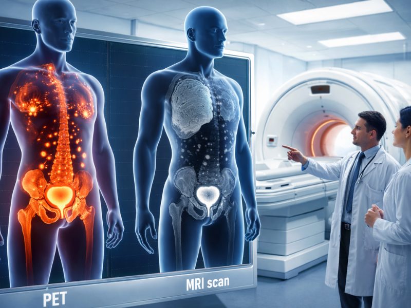
In a phase 2 prospective trial (Next Generation Trial; ClinicalTrials.gov identifier: NCT05141760) conducted across two hospitals in Alberta, Canada, researchers found that a new imaging technique called 18F-PSMA-1007 PET/CT significantly outperforms the current standard, multiparametric MRI, in accurately staging prostate cancer before surgery. The study demonstrated that PSMA PET is superior in identifying the correct tumor stage, detecting the dominant tumor nodule, determining cancer laterality, and spotting extracapsular extension.1
According to the authors, these results suggest that 18F-PSMA-1007 PET/CT could become an essential tool in the preoperative workflow for patients with prostate cancer, potentially improving treatment planning for men with intermediate- and high-risk prostate cancer.
“18F-PSMA-1007 was correct approximately twice as often as MRI when identifying tumor stage,” said corresponding author Adam Kinnaird, MD, PhD, who is an assistant professor at the University of Alberta. “Our findings will give patients more information about their disease prior to treatment, helping them to choose the ideal treatment option for them.”
The report was published in JAMA Oncology.
Need for Accurate Preoperative Imaging Methods for Prostate Cancer
Accurate staging of prostate cancer is crucial for determining the most appropriate treatment strategy and predicting patient outcomes. Currently, multiparametric MRI is the recommended imaging technique for the diagnosis and locoregional staging of prostate cancer before radical prostatectomy.1 However, MRI has limitations in detecting extracapsular extension and accurately determining the full extent of the disease.
Commenting on their rationale for comparing 18F-PSMA-1007 PET/CT with multiparametric MRI, Dr. Kinnaird explained that recent studies have shown that prostate-specific membrane antigen (PSMA) PET is superior to conventional imaging for metastatic staging of prostate cancer. However, studies on second-generation PSMA PET radioligands for locoregional staging have been limited. This knowledge gap led researchers to investigate whether 18F-PSMA-1007 PET/CT could provide more accurate staging information than the current standard of care.
“18F-PSMA-1007 is a new radioligand that has minimal urinary excretion,” Dr. Kinnaird said. “Therefore, we hypothesized it would be more accurate than conventional radioligands that are excreted in the urine for tumor localization in the prostate gland.”
A Phase 2 Trial Comparing 18F-PSMA-1007 to Multiparametric MRI
The Next Generation Trial was designed as a phase 2 prospective validating paired cohort study. The researchers recruited 150 men with intermediate-risk or high-risk prostate cancer who were considering robot-assisted laparoscopic radical prostatectomy.1 These patients underwent both 18F-PSMA-1007 PET/CT and multiparametric MRI within two weeks of each other and at least five days before surgery.
To reduce the risk of bias, radiologists, nuclear medicine physicians, and pathologists were blinded to all other imaging, biopsy, and clinical data for each participant.1 The MRI scans were interpreted using the Prostate Imaging Reporting and Data System (PI-RADS) version 2.1 criteria, and the PET scans were interpreted using modified Prostate Cancer Molecular Imaging Standardized Evaluation (PROMISE) guidelines.1
The primary outcome of the study was the correct identification of the prostate tumor stage by each imaging test. Secondary outcomes included correct identification of the dominant nodule, laterality, extracapsular extension, and seminal vesicle invasion.1 The final analysis included 134 patients who underwent radical prostatectomy, with histopathological results serving as the reference standard.
PSMA PET Outperforms MRI in Prostate Cancer Staging
PSMA PET significantly outperformed MRI in accurately identifying the pathological tumor stage, with 45% accuracy compared to 28% for MRI (P = 0.003).1 This means that PSMA PET correctly staged the cancer in nearly half of the cases, whereas MRI was accurate in just over a quarter of the cases.
PSMA PET also demonstrated superiority in several other aspects of cancer assessment, including dominant nodule identification, laterality determination, and extracapsular extension detection. PSMA PET correctly identified the dominant tumor nodule in 94% of cases, compared to 83% for MRI (P = 0.01). Moreover, PSMA PET accurately determined whether the cancer was unilateral or bilateral in 64% of cases versus 44% for MRI (P = 0.001). PSMA PET also identified extracapsular extension (cancer spreading beyond the prostate capsule) in 75% of cases, compared to 63% for MRI (P = 0.01). Although not statistically significant, PSMA PET showed a trend toward better detection of seminal vesicle invasion (91% versus 85%, P = 0.07).
Importantly, PSMA PET demonstrated higher sensitivity for extracapsular extension (58% versus 33%) than MRI, while maintaining similar specificity (~90%).1 This means that PSMA PET was better at detecting when cancer had spread beyond the prostate without increasing false positives.
In addition, PSMA PET detected more clinically significant prostate cancer nodules (Gleason grade group 2 or higher) than MRI (87% versus 62%, P < 0.001).1 According to the authors, this improved detection rate could have significant implications for treatment planning and patient outcomes.
Commenting on the implications of these findings, Dr. Kinnaird said, “Our results suggest that men should have 18F-PSMA-1007 PET/CT prior to radical prostatectomy if nerve sparing is planned or prior to focal therapy to ensure that the disease is unilateral.”
Limitations and Future Work
The authors acknowledged that the study has limitations, including the lack of whole-mount specimens for pathological analysis, which could have provided even more detailed correlations between imaging and histopathology.1 Furthermore, the lack of a standardized extended template for lymph node dissection during radical prostatectomy limited the ability to determine the accuracy of nodal staging for each imaging type.1
“The readers of the imaging were blinded to other scans, biopsy information, and clinical parameters, which was important for the fidelity of the trial. However, in real-world practice, a nuclear medicine physician or a radiologist would be able to incorporate these results into their interpretation of the images,” noted Dr. Kinnaird.
Commenting on their plans to further explore the utility of PET/CT in prostate cancer diagnosis, Dr. Kinnaird said:
“We have now started a multicenter randomized controlled trial called PSMA-Guided Ablation of the Prostate that uses 18F-PSMA-1007 PET/CT to determine eligibility for focal therapy.”
The team also plans to perform a secondary analysis of the trial in which nuclear medicine physicians and radiologists hold multidisciplinary rounds and are unblinded to both the PET/CT and the MRI findings, using the information from both to determine the tumor stage.
The study received financial support from the Canadian Urological Association, the University Hospital Foundation, and the Royal Alexandra Hospital Foundation.
References
- Mookerji N, Pfanner T, Hui A, et al. Fluorine-18 Prostate-Specific Membrane Antigen-1007 PET/CT vs Multiparametric MRI for Locoregional Staging of Prostate Cancer. JAMA Oncol. 2024;10(8):1097-1103. doi:10.1001/jamaoncol.2024.3196